&geometry(278x56) "Veterinary Specialist Services Pty Ltd.")
Treatment of Cruciate Ligament Tears in Dogs
What is a cruciate ligament?
Dogs have similarities to people in how their joints are stabilised and move. In people, the anterior cruciate ligament (ACL) is a common cause of joint problems in the knee. It is typically damaged in an active activity and is common in sportspeople performing movements that require rapid stopping or changes of direction. In dogs, there is a ligament in the stifle (knee joint) that performs a very similar function - it is called the cranial cruciate ligament.
How does the cranial cruciate ligament become damaged in dogs?
In most dogs, the ligament begins to deteriorate over time. There have been many investigations into the underlying reasons for this, but the causes are not completely clear. The ligament typically partially tears over time, and as the tear worsens, so does the degree of lameness. It is still possible to get an acute complete tear while the dog is undertaking vigorous activity like chasing a ball or running at the beach. For many of these dogs, once they have x-rays, it becomes apparent that there has been some issue with the joint prior to the activity that seemingly led to the damage.
What else can happen when a dog tears the cranial cruciate ligament?
There are other structures in the stifle (knee) joint that help to stabilise the joint and distribute pressure between the tibia (shin bone) and femur (thigh bone). The most important of these are known as the menisci. The meniscus on the inside of the joint (medial meniscus) is the most frequently damaged cartilage and is more likely to be damaged with a complete tear of the cranial cruciate ligament.
How is a dog assessed for treatment of cranial cruciate ligament problems?
A consultation and physical exam are the first steps in patient assessment. Typically, a discussion is held, to try to gain information from the family about the symptoms they have noticed and how these have progressed, as well as ensuring that the general health of the patient is OK. A veterinarian will often what the dog to determine which legs appear to show any signs of problems. Around 20% of dogs with cranial cruciate ligament problems will have both hind legs affected when they are assessed by the vet. The vet will then palpate the knee and apply pressure in various directions to determine if the ligament has been damaged. Following this, it is typical that x-rays are advised. These perform a few functions, including ruling out other causes of lameness, ensuring that the typical radiographic (x-ray) signs of cruciate ligament disease are present and, in some cases, aid in planning for surgery. It is not possible to see the cruciate ligament on an x-ray, so the diagnosis is presumptive based on both palpation and x-rays.
What are the options for treatment of a cruciate ligament tear in dogs?
Treatment for cranial cruciate ligament disease is typically surgical. An older study suggests that around 20% of dogs will manage with medical management alone. Non-surgical treatment may also be occasionally advised, if there are other significant health issues that mean an anaesthetic is not in the patient’s best interest. The reality is that most dogs develop progressive arthritis with untreated cranial cruciate ligament disease. Regardless of which type of definitive surgery is selected, all patients should have the inside of their joint examined as part of the surgery. This allows a few things to happen, the most important being confirmation that the ligament is torn and assessing and treating any damage to the meniscus cartilages. Lack of treatment of a meniscus cartilage injury typically leads to ongoing lameness after surgery. Currently, removal of the damaged portion of the meniscus is the only real option for management. The best way to assess the joint is through arthroscopy. This is where a camera and small instruments are used to examine the joint and treat any problems with the meniscus cartilage. This approach provides better visualisation (compared to opening the joint up completely) and a faster recovery. Arthroscopy is typically only available in referral surgical centres, such as Veterinary Specialist Services, and requires specific training.
There are a few surgical options available. The treatment for human patients is to use some of a hamstring tendon to replicate the ligament. Over a 9-to-12-month period, with good rehabilitation, this can become similarly effective to the native ligament. Unfortunately, this approach is not reliably successful in dogs, mostly due to the difficulty in confinement and rehabilitation compared to people. The most commonly recommended surgical option for most patients is a Tibial Plateau Levelling Osteotomy (TPLO) or Triple Tibial Osteotomy (TTO). These surgical procedures aim to remove the need for the ligament by changing the way the joint functions. There are many publications investigating surgical options for treatment of cranial cruciate ligament disease in dogs. The best quality publications support the use of TPLO surgery as the best option for most dogs, and a very high proportion of patients (>90%) will return to athletic active function. TPLO surgery can be performed on dogs of a wide range of sizes (we have treated dogs who weigh from 2kg to >100kg with this procedure). For some patients who are less than 15kg in body weight, a prosthetic ligament alternative that is placed around the outside of the joint (known as an extra capsular suture) is used.
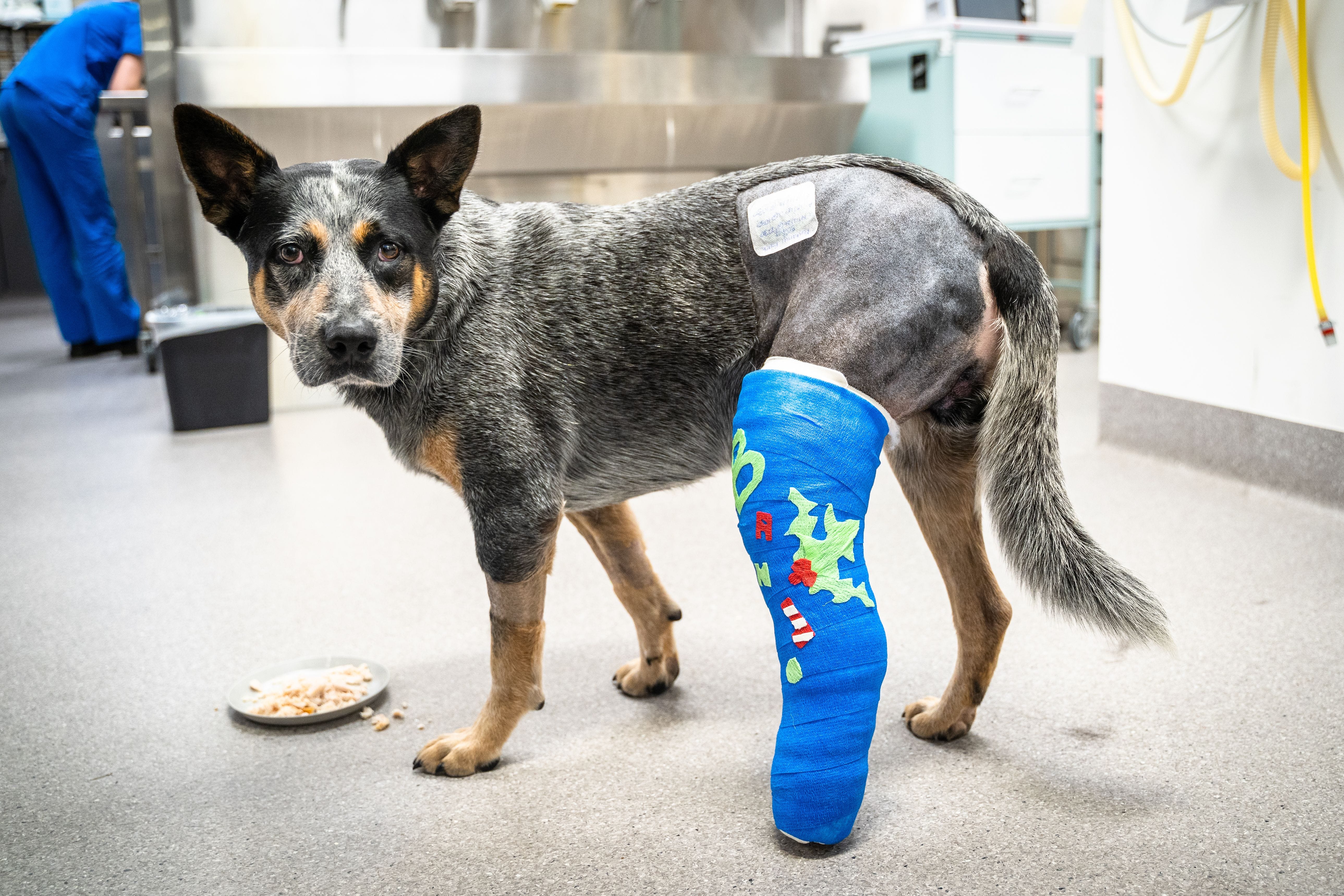
What should we expect after surgery?
The initial post-operative period is typically focused on pain management and recovery from the procedure. Most dogs will have multiple pain medications and sometimes antibiotics. Patients need to be confined for at least 6 weeks (but sometimes up to 12 weeks) after surgery to reduce the risk of damage to the surgery. It is important that the patient walks on the leg, as this helps the recovery process. Most dogs will be walking consistently on the leg with an obvious limp by 2 weeks post-surgery and walking with a mild limp by 6 weeks post-surgery. Around 80% of the improvement after surgery occurs within the first 12 weeks, with the remainder taking the rest of a calendar year. Most dogs return to a high level of athletic activity, but osteoarthritis is always possible. A structured physiotherapy program has been shown to improve the rate of recovery, as has a comprehensive approach to arthritis management. Most family vets have a considered approach to arthritis management. A plan should be made with the family to watch out for signs of complications. The most common complications of this type of surgery are bacterial infection, subsequent damage to the meniscus cartilage or damage to the surgical repair, if the patient is too active following surgery. Most complications can still be managed to achieve a successful result for the patient.
For more information on the treatment and management of cruciate ligament tears in dogs or for advice on specific cases, please contact our surgery team on 1800 442 648.
| Tags:Emergency/Critical CareNewsOrthopaedicsPatient Care |