"Veterinary Specialist Services Pty Ltd.")
Case Study: Oesophageal Foreign Body Obstruction
 "Case Study: Oesophageal Foreign Body Obstruction")
Oesophageal Foreign Body Obstruction, Thoracotomy, Oesophageal Resection and Anastomosis
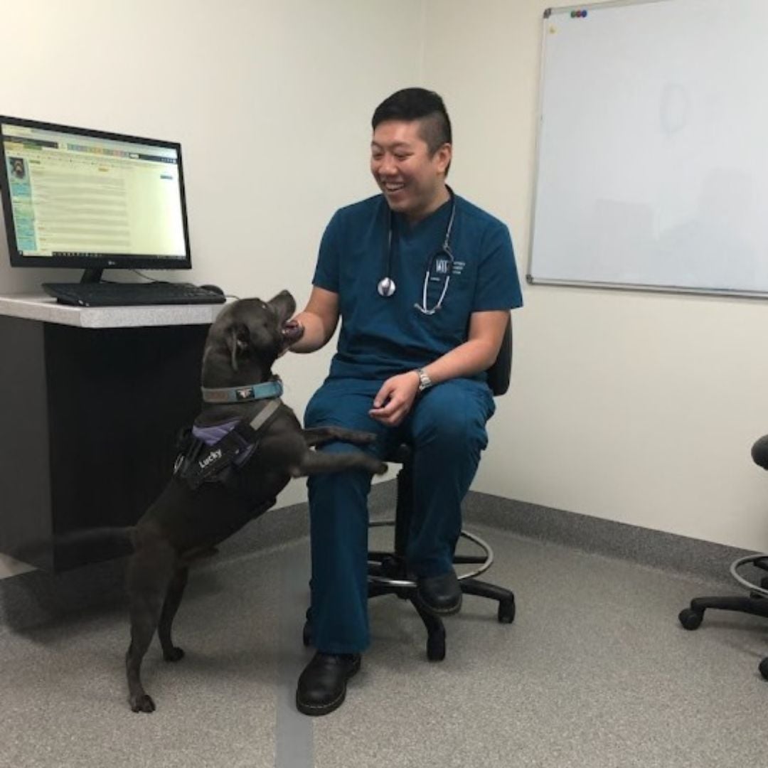
One year ago, Lucky, a three-year-old Staffy, was transferred to Specialist Surgeon Dr Jason Hoon for intervention of a perforating oesophageal foreign body obstruction.
Lucky had been to his family vet clinics (Manly Road Vet and Koala Park Vet Clinic) for the onset of symptoms that included vomiting, twitching, inappetence and just not seeming himself. On investigation, an obstructive oesophageal bone foreign body was diagnosed and he was referred to Animal Emergency Service (AES) for intervention. Endoscope evaluation by the AES team identified severe necrosis of the oesophagus and perforation, due to the bone foreign body, requiring emergency surgery.
AES then transferred Lucky to the care of VSS for further work-up and surgery. Due to the tricky location affected within the chest cavity, Dr Hoon performed a thoracotomy (open chest surgery) to allow for a 4cm oesophageal resection and anastomosis. This is a procedure performed in dogs to remove a section of the diseased oesophageal tract and reconstruct it, keeping the remaining healthy parts intact. A gastrostomy tube (G-Tube) was also placed so Lucky could get nutrition via a feeding tube directly into his stomach for a week post-operatively. Lucky remained in PetICU for his recovery journey and was slowly re-introduced to oral feeding via diet trials to ensure safe healing of the oesophagus.
Lucky was certainly a very lucky boy - he has continued to maintain an excellent recovery one year on from this terrible incident, especially considering his complex surgical journey.
| Tags:Diagnostic TestingEmergency/Critical CareGastrointestinalNewsPatient CareSurgery |